Three Tools to a Great Root Canal Experience for your Patients

When patients are referred to us for endodontic treatment, their dentists are counting on a favorable outcome. At Elm Endodontics, our goal is not only to deliver a favorable outcome, but a favorable experience. Root canal treatment gets a bad rap, when in reality it can be a relatively good experience when done in the right environment. Three tools in our office that improve the experience for the patient are the dental operating microscope (DOM), the cone beam CT (CBCT), and our website – all of which allow us to communicate better with our patients and our doctors.
The Dental Operating Microscope
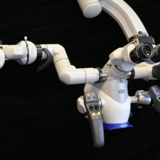
The dental operating microscope (DOM) has been around in the field of dentistry since the eighties, and has grown slowly in popularity to this day. It is used by many endodontists and by some dentists. Endodontists vary in the degree that they use the DOM; some use it on every case from start to finish, and others use it only when they think they need it. The DOM improves the treatment experience in at least four ways.
The first way that it improves the treatment experience is that it offers improved ergonomics for the doctor. This is important so that by the end of the day the doctor is less fatigued and will be able to perform as well on the last case as he did on his first. It also allows for consistency in treatment procedure and quality.
The second way that the DOM improves the treatment experience (when an assistant scope is attached) is that the assistant can be more engaged in the treatment. This allows the assistant to be quicker to deliver care like suctioning or passing instruments, ultimately resulting in a more efficient, and thus quicker, appointment for the patient.
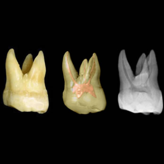
The third reason why the DOM improves the endodontic experience is that it allows the doctor to see and treat the finer details of the case that may affect the long-term success of the root canal treatment. Obstacles such as calcifications (pulp stones) may be present in the root canal space that may not even be seen if the endodontist is not using a microscope, and thus may block the canal in a way that makes it impossible to fill completely. When I do a root canal procedure and the case is nearing completion, I will pause and inspect the inside of the tooth under high magnification. Often I will find remaining pulp (nerve) tissue along the walls. This tissue can be easily removed when I am aware of it, but would otherwise remain in the tooth if I didn’t use the microscope for close inspection. Dental loupes (the magnifiers typically worn by dentists) don’t provide enough magnification and lighting to see these fine details. Leaving tissue in the tooth provides bacteria with a nutrition source and may contribute to failure of the root canal many years later. Keep following this blog where I will post more examples of how the microscope helps in treatment.
The fourth reason why the dental operating microscope improves the root canal experience is that it can easily allow the doctor to take pictures of the case and to share that information with the patient or doctor. Taking a picture of the patient’s actual cracked tooth or periodontal probing and using that picture in discussion with the patient makes for a more powerful teaching tool than using canned, pre-fabricated drawings or images. In addition, it improves communication with the referring dentist. I have on many occasions emailed a photograph of a patient sitting in my chair, called the dentist, and made treatment planning decisions over the phone that expedited treatment for the patient, allowing them to get immediate treatment rather than waiting for time-consuming exchange of information and decision making.

The Cone Beam CT (Computed Tomography)
This radiographic tool has entered the field of endodontics only in the last couple of years. Very few endodontists have one, but the number of those who do is slowly growing. At the time of this writing I am aware of only about a half dozen endodontists in the Denver area that have a CBCT. This tool allows us to image an area of the patient’s mouth in three dimensions, allowing us to see the tooth from a minimum of three sides including the top. Traditionally the endodontist would point to a small radiograph on a light box and try to explain what was going on, and the patient would just have to take the doctor at his word. Then the last decade brought us digital radiography that allowed us to display the tooth on a computer screen. This greatly improved doctor-patient communication because the patient could now see what the dentist was pointing at. For years I would use this tool and try to verbally explain to the patient how the two dimensional image represented their three dimensional tooth. Although patients would nod and agree, I didn’t feel like they truly understood what I was trying to explain.
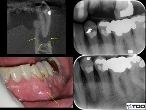
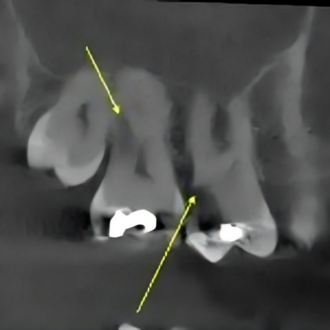
With the CBCT, I can now show the patient’s actual tooth from the front, side, or top view and show the actual missed canal, infection into the sinus, or many other scenarios. Because the areas of infection are represented more clearly, I can see in their faces that they understand more clearly why I am recommending treatment. The CBCT therefore not only helps to improve the delivery of care, but it acts as a great communication tool to explain endodontic treatment.


Our Website
The third tool that helps to improve the patient’s endodontic experience is our website. Although many patients welcome a referral to a specialist, many are a little apprehensive to get treatment from someone new. I understand that fear and anxiety in dentistry stem primarily from a lack of trust. I have designed our website so that patients can get to know us and our procedures before they arrive. I feel that this tool will remove some of the mystery behind the referral process and specialty care. Patients can also register on our website which will reduce the time they spend addressing administrative items when they arrive. If we put this amount of effort into our website, imagine the effort we put into our actual treatment.
We have added advanced technology like the microscope and the cone beam CT to our office to deliver both simple and complex treatment in an efficient manner. The primary goal is to deliver high quality care, but a great benefit is that it improves patient education, reduces anxiety, and creates a better experience for the patient.


